|
|
Calcaneal Fractures
Lover's Fracture
General Considerations
- Calcaneous is the most commonly fractured tarsal bone
- A so-called “lover’s Fracture” is an intra-articular fracture produced by an axial loading force typically produced by a leap from a height with person landing on heels (also called a “Don Juan” fracture)
- Why is it called a “Lover’s fracture?”
- Because it is the type of fracture that could presumably be caused by a lover jumping out of the bedroom window to escape from a surprised and enraged spouse
- Bilateral in up to 10%
- The same axial load is transmitted to the spine and may produce associated burst fractures of the lumbar or thoracic spine in up to 10% of patients
- Frequently occur at the thoraco-lumbar junction at L1, L2, T12 and T11
Classifications
- Intra-articular (25%)
- More common in diabetics
- Recover function well
- Intra-articular (75%)
- Stress fractures of the calcaneous

- Uncommon overall
- Associated with osteoporosis
- Also occur with repetitive stress, as in runners
- Conventional radiographs may not show the fracture until healing is occurring
- Bone scan or MRI will be more sensitive
- Sander’s classification based on CT findings
Sander’s classification based on CT findings |
Type 1 |
Non-displaced |
Type 2 |
Split in 2 parts |
Type 3 |
3 part with depression of posterior facet |
Type 4 |
Severely comminuted |
Clinical findings
- Pain
- Swelling
- Deformity following trauma
Imaging Findings
- Conventional radiographs are the study of first choice
- Lateral and axial views
- Radiographs of the thoraco-lumbar spine may also be indicated
- CT is the examination of choice to demonstrate all of the fragments and their relationships
- Boehler's angle is measured at the intersection of a line drawn between the posterior superior aspect of the calcaneal tuberosity to the highest point of the posterior articular facet and another line drawn to the anterior process of the calcaneous
- It is normally 25-40°
- As the calcaneous compresses secondary to a fracture, there will be a decrease in Boehler’s angle to less than 20° (frequently to 0°)
Treatment
- Extra-articular fractures are usually managed conservatively without surgery
- The treatment of intra-articular fractures is controversial
- Up to 10% with compression-type fracture are associated with compartment syndrome
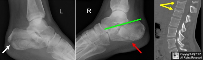
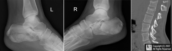
Bilateral calcaneal fractures and fractures of spine. There is a comminuted fracture of the left calcaneous (white arrow); there is a comminuted fracture of the left calcaneous (red arrow) with flattening of Boehler's Angle to 0 degrees. A sagittal reconstructed CT scan of the spine shows compression fractures of the superior endplates of T12 and L1 (yellow arrows)
.
For additional information about this disease, click on this icon above.
For this same photo without the arrows, click here
|
|
|

)
)